- UHR imaging of beam-sensitive samples at low-kV
- Routine investigation of heavy-metal stained resin-embedded samples with excellent contrast using the In-beam BSE detector at low-kV
- Best resolution Ga FIB column with optimized ion optics for excellent performance and reproducibility
- Site-specific TEM lamella preparation and in-situ visualization with an easy-to-use STEM detector that offers cost-effective imaging results comparable to TEM
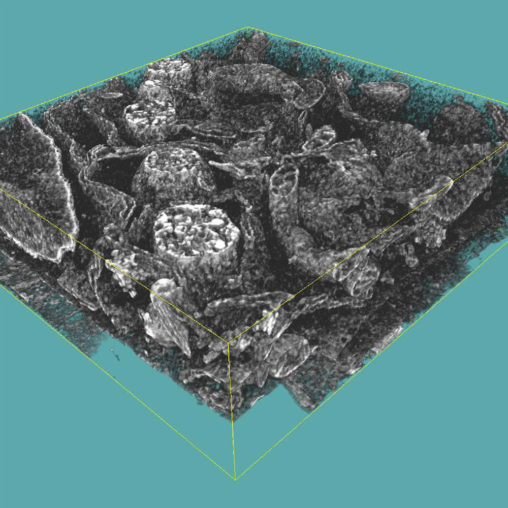
- 3D nanovisualization and volume reconstruction of resin-embedded cellular structures
- Cryo UHR SEM imaging and cryo-FIB-SEM applications
-

-
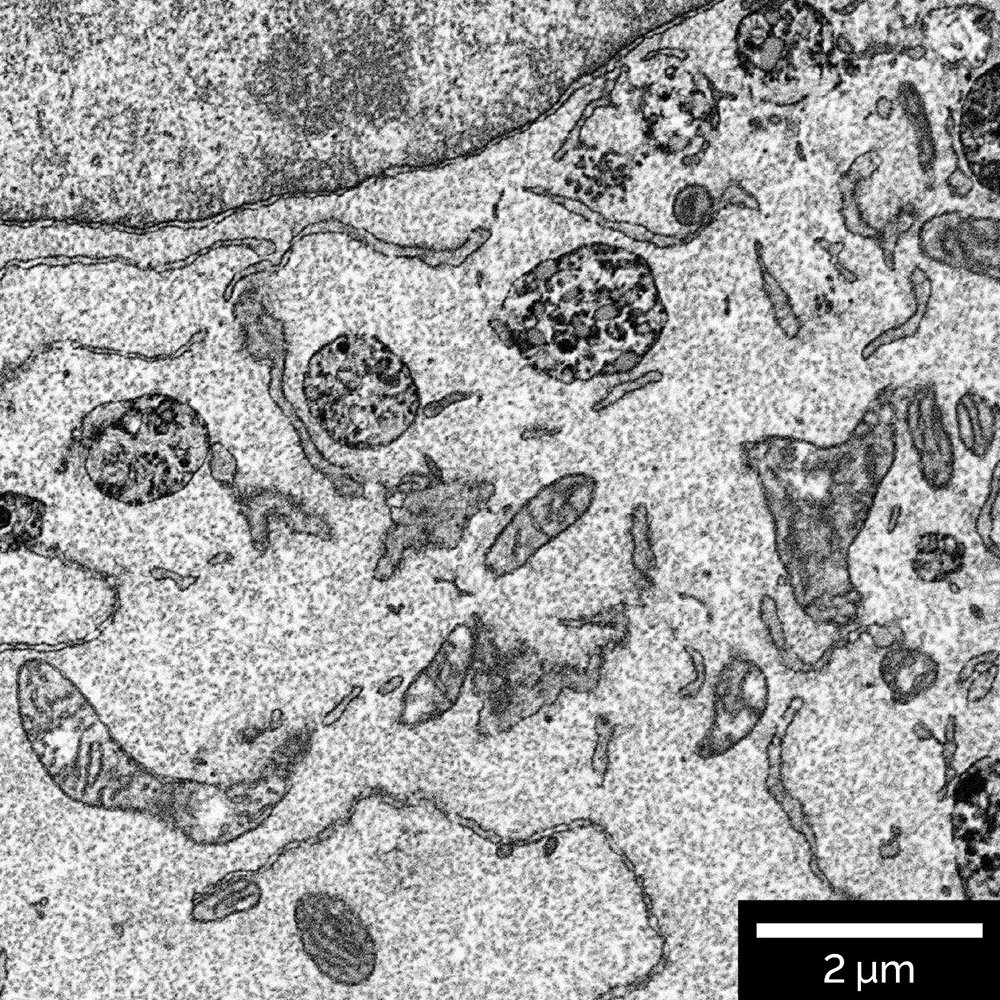
Resin embedded mammalian cells. Additionally inverted image was acquired using In-Beam BSE detector at accelerating voltage 2.5 keV
-

-
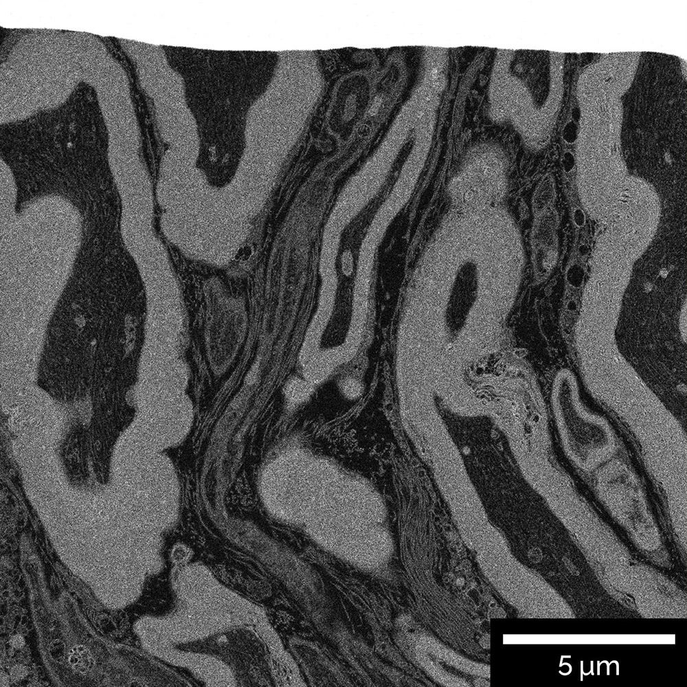
Resin embedded brain tissue. Cross-section was prepared with Ga FIB and visualized using In-Beam BSE detector
-

-
3D ultrastructural reconstruction of mammalian cell at 5 × 5 × 8 nm voxel size sampling in x, y, and z dimensions. Selected FIBSEM stack shows intracellular organization inside the cell


